- Microscope Manuals Pdf
- Science Microscope Kit
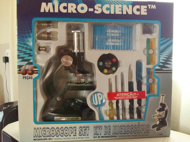
Jan 30, 2012 MICRO SCIENCE BEST MICROSCOPE SET FOR 8 Jan 2018 A wide variety of micro science microscope options are available to you, such as Well I have the instruction manual, but it is not too helpful. Deluxe Microscope Set By Soma -This 84 piece set includes (1) metal die cast28 Oct 2014 2. The MicroChem Kit Standard Edition includes a 119 page MicroChem Kit Manual which explains each experiment covering the following: - Goals The goals section outlines the major learning areas of the experiment.
Do you need help learning how to effectively use a compound microscope?
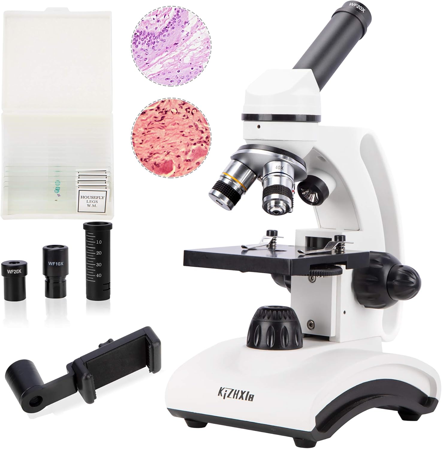
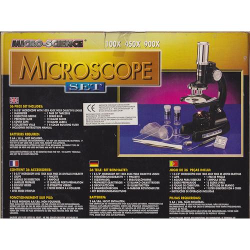
Eastcolight Microscope Set comes with 8.625' Microscope with 100X, 450X, 900X, Micro-Science Data Bank,Prepared Slides, Blank Slides, Cover Glasses, Slide Covers, Blank Labels, Collecting Vials, Plastic Cylinders, Lens Cap, Stirring Rod, Petri Dish, Hatchery (Micro-Incubator), Instruction Manual, 2 AA Batteries required (Not included). Instructions, manuals, brochures and catalogues. If you buy a used microscope, it probably won’t be supplied with an instruction manual, and most manufacturers don’t keep copies of manuals for discontinued microscopes. Fortunately, there are several websites where enthusiasts have collected PDFs of manuals that you can download.

It’s not as complicated as it may look – in fact, we hope these instructions will help you quickly become comfortable and proficient at using your microscope!
Find a great explanation of compound and stereo microscopes; definitions of the terms and parts of a microscope, and a great selection of quality microscopes on our Microscopes page.
How to Use Your Compound Microscope |
- Set your microscope on a tabletop or other flat, sturdy surface where you will have plenty of room to work. Plug the microscope’s power cord into an outlet. (Note: some compound microscopes don’t use electric lighting, but have a mirror to focus natural light instead.)
- Switch on your microscope’s light source and then adjust the diaphragm to the largest hole diameter, allowing the greatest amount of light through. If you have an iris diaphragm, slide the lever till the most light comes through. See the diagram below for help locating these parts.
- Rotate the nosepiece to the lowest-power objective usually 4x for 40x magnification). It is easiest to scan a slide at a low setting, since you have a wider field of view at low power.
- Place a microscope slide on the stage, either under the stage clips or clipped onto the mechanical stage if your microscope has one. A prepared slide works best when you do this for the first time. (If you do not have a prepared slide, place a strand of colored yarn or thread on a blank slide and place a coverslip over it.) Move the slide until the specimen is under the objective lens.
- Adjust the large coarse focus knob until the specimen is in focus. Slowly move the slide to center the specimen under the lens, if necessary. Do this by nudging it gently with your fingers or by turning the slide control knobs if you have a mechanical stage.
- Adjust the small fine focus knob until the specimen is clearly in focus. Then adjust the diaphragm to get the best lighting. Start with the most light and gradually lessen it until the specimen image has clear, sharp contrast.
- Scan the slide (right to left and top to bottom) at low power to get an overview of the specimen. Then center the part of the specimen you want to view at higher power.
- Rotate the nosepiece to the 10x objective for 100x magnification. Refocus and view your specimen carefully. Adjust the lighting again until the image is most clear (you will need more light for higher power). Repeat with the 40x objective for 400x magnification, which will enable you to see all of the specimen detail that’s necessary for high school biology lab work.
- Optional: If your microscope has a 100x oil-immersion lens, you’ll need to put 1-2 drops of immersion oil over the slide coverslip (the piece of glass over the middle of the slide) before viewing it at the highest power. Move the 100x objective lens into position, and then slowly move the stage up until the lens makes contact with the oil. Continue focusing with the coarse focus knob until the color or blurred outline of the specimen appears. Finish focusing with the fine focus knob. With the 100x lens, you will be able to see additional cell detail, but you will need to take extra care with focus and contrast for a clear image. When you are done using the slide, clean the oil off of the slide and the lens with lens cleaning paper and solution.
Once you’ve mastered the basics of using your microscope, you can expand your microscope studies. Print out copies of our Microscope Observation worksheet to help you record what you see! |
Diagram of Microscope Parts |
Microscope Cleaning and Maintenance |
| To clean the exterior side of lenses, use a non-solvent cleaning solution designed for cleaning optics or eyeglasses. First remove dust with a soft brush or can of compressed air. Then moisten a piece of dry lint-free tissue or lens paper and clean the lens surfaces with a circular motion. Repeat with a second piece of paper moistened with lens solution if necessary. Repeat once again with dry lens paper until the lens is clean and dry. Use this same procedure for the eyepiece and objective lenses. To clean the interior side of lenses, DO NOT use the above procedure. Rather use a bulb-type duster or a compressed gas canister designed specifically for cameras/microscope cameras and other optical equipment. Only use a microfiber cloth. Order our Microscope Cleaning Kit for a complete set of tools to clean your microscope with. To clean the body of the microscope, use a soft dry or damp cloth. Using a dust cover will help keep your microscope investment clean and dust-free. |
More Information
Find an affordable set of prepared slides to use with your microscope, whether you’re interested in anatomy, zoology, botany, or just want a starter set.

Check out our tips for making slides and doing other activities with your microscope.


Do you need help learning how to effectively use a compound microscope?
Microscope Manuals Pdf
It’s not as complicated as it may look – in fact, we hope these instructions will help you quickly become comfortable and proficient at using your microscope!
Science Microscope Kit
Find a great explanation of compound and stereo microscopes; definitions of the terms and parts of a microscope, and a great selection of quality microscopes on our Microscopes page.

How to Use Your Compound Microscope |
- Set your microscope on a tabletop or other flat, sturdy surface where you will have plenty of room to work. Plug the microscope’s power cord into an outlet. (Note: some compound microscopes don’t use electric lighting, but have a mirror to focus natural light instead.)
- Switch on your microscope’s light source and then adjust the diaphragm to the largest hole diameter, allowing the greatest amount of light through. If you have an iris diaphragm, slide the lever till the most light comes through. See the diagram below for help locating these parts.
- Rotate the nosepiece to the lowest-power objective usually 4x for 40x magnification). It is easiest to scan a slide at a low setting, since you have a wider field of view at low power.
- Place a microscope slide on the stage, either under the stage clips or clipped onto the mechanical stage if your microscope has one. A prepared slide works best when you do this for the first time. (If you do not have a prepared slide, place a strand of colored yarn or thread on a blank slide and place a coverslip over it.) Move the slide until the specimen is under the objective lens.
- Adjust the large coarse focus knob until the specimen is in focus. Slowly move the slide to center the specimen under the lens, if necessary. Do this by nudging it gently with your fingers or by turning the slide control knobs if you have a mechanical stage.
- Adjust the small fine focus knob until the specimen is clearly in focus. Then adjust the diaphragm to get the best lighting. Start with the most light and gradually lessen it until the specimen image has clear, sharp contrast.
- Scan the slide (right to left and top to bottom) at low power to get an overview of the specimen. Then center the part of the specimen you want to view at higher power.
- Rotate the nosepiece to the 10x objective for 100x magnification. Refocus and view your specimen carefully. Adjust the lighting again until the image is most clear (you will need more light for higher power). Repeat with the 40x objective for 400x magnification, which will enable you to see all of the specimen detail that’s necessary for high school biology lab work.
- Optional: If your microscope has a 100x oil-immersion lens, you’ll need to put 1-2 drops of immersion oil over the slide coverslip (the piece of glass over the middle of the slide) before viewing it at the highest power. Move the 100x objective lens into position, and then slowly move the stage up until the lens makes contact with the oil. Continue focusing with the coarse focus knob until the color or blurred outline of the specimen appears. Finish focusing with the fine focus knob. With the 100x lens, you will be able to see additional cell detail, but you will need to take extra care with focus and contrast for a clear image. When you are done using the slide, clean the oil off of the slide and the lens with lens cleaning paper and solution.
Once you’ve mastered the basics of using your microscope, you can expand your microscope studies. Print out copies of our Microscope Observation worksheet to help you record what you see! |
Diagram of Microscope Parts |
Microscope Cleaning and Maintenance |
| To clean the exterior side of lenses, use a non-solvent cleaning solution designed for cleaning optics or eyeglasses. First remove dust with a soft brush or can of compressed air. Then moisten a piece of dry lint-free tissue or lens paper and clean the lens surfaces with a circular motion. Repeat with a second piece of paper moistened with lens solution if necessary. Repeat once again with dry lens paper until the lens is clean and dry. Use this same procedure for the eyepiece and objective lenses. To clean the interior side of lenses, DO NOT use the above procedure. Rather use a bulb-type duster or a compressed gas canister designed specifically for cameras/microscope cameras and other optical equipment. Only use a microfiber cloth. Order our Microscope Cleaning Kit for a complete set of tools to clean your microscope with. To clean the body of the microscope, use a soft dry or damp cloth. Using a dust cover will help keep your microscope investment clean and dust-free. |
More Information
Find an affordable set of prepared slides to use with your microscope, whether you’re interested in anatomy, zoology, botany, or just want a starter set.
Check out our tips for making slides and doing other activities with your microscope.


